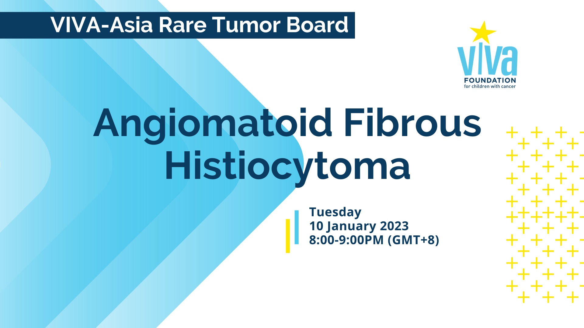
VIVA-ASIA RARE TUMOR BOARD WEBINAR is a virtual quarterly meeting that is organized by VIVA Foundation for Children with Cancer to enable doctors from all over the world to discuss the diagnosis and treatment of rare pediatric tumours.
Join us for a discussion about Angiomatoid Fibrous Histiocytoma (AFH). A team from The Hong Kong University – Shenzhen Hospital in Hong Kong will present on Angiomatoid Fibrous Histiocytoma. Dr Debbie Chong from KK Women’s and Children’s Hospital (KKH) in Singapore will share the team’s experience by reviewing a series of cases, while Dr Kenneth Chang, Head of the Department of Pathology and Laboratory Medicine at KKH, will discuss the pathology and molecular pathology of AFH.
If you can’t make it, you can still register to receive a copy of the webinar recording.
Medical professionals in Singapore may eligible for CME/CPE/CNE credits for attending this webinar.
Credits and Certificates of Attendance will only be issued for those who are able to attend the live session and are in adherence with VIVA’s Attendance Policy.
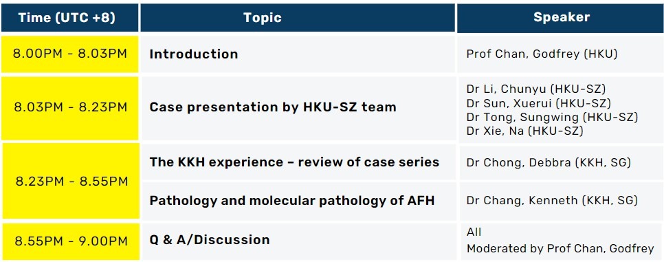
PROGRAMME RUNDOWN
SYNOPSIS
Case Presentation by HKU-SZ Team
A 12-year-old Chinese boy presented with a painful right axillary mass with multiple enlarged lymph nodes. MRI examination was performed in another hospital, and the diagnosis was synovial sarcoma. The diagnosis of pathological examination combined with NGS suggested AFH. After 2 cycles chemotherapy, PET-CT showed a hypermetabolic nodule in liver, while MRI revealed multiple nodules in liver. MRI examination was rechecked one month later, and no lesions were found in the liver. A 12-year-old Chinese boy presented with a painful right axillary mass with multiple enlarged lymph nodes. MRI examination was performed in another hospital, and the diagnosis was synovial sarcoma. The diagnosis of pathological examination combined with NGS after tumor resection suggested Angiomatoid Fibrous Histiocytoma. After 2 cycles chemotherapy, PET-CT showed a hypermetabolic nodule in liver, while MRI revealed multiple nodules in liver. MRI examination was rechecked one month later, and no lesions were found in the liver.
WATCH IT AGAIN!
PRESENTERS
Dr Li, Chunyu
Senior Medical Officer The University of Hong Kong – Shenzhen Hospital Hong Kong
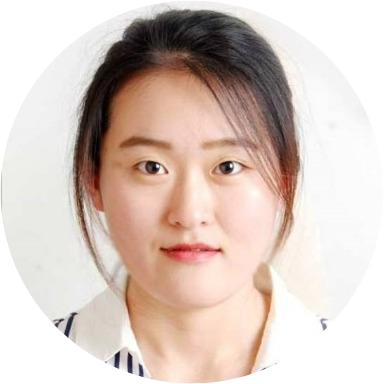
Dr Sun, Xuerui
Resident, Department of Pathology , The University of Hong Kong – Shenzhen Hospital, Hong Kong
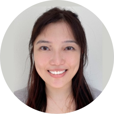
Dr Xie, Na
Consultant Radiologist, Department of Radiology, The University of Hong Kong – Shenzhen Hospital Hong Kong
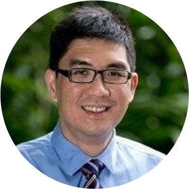
Dr Chang, Kenneth
Head and Senior Consultant Department of Pathology and Laboratory Medicine, KK Women’s and Children’s Hospital, Singapore
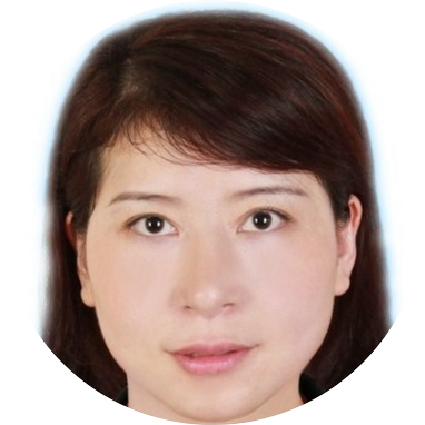
Dr Chong, Debbra
Consultant, Haematology-Oncology Service KK Women’s and Children’s Hospital, Singapore
CONVENORS
Prof Chan, Godfrey
Honorary Professor, Department of Paediatrics and Adolescent Medicine School of Clinical Medicine, The University of Hong Kong Consultant Paediatric Haematologist Oncologist Hong Kong Sanatorium and Hospital Hong Kong SAR
Dr Liu, Anthony
Honorary Clinical Assistant Professor Department of Paediatrics & Adolescent Medicine LKS Faculty of Medicine, The University of Hong Kong Hong Kong SAR
Staff Oncologist, Pediatric Brain Tumour Program The Hospital for Sick Children, Toronto Canada